

67% less
re-op
Up to 67% reduction in re-operation rates 1

81-100% sensitivity
Demonstrated across clinical studies 1,2,3

92-99% specificity
Demonstrated across clinical studies 1,2,3

Clinician
assessment
Images interpreted accurately by surgeons & pathologists 1,2,3
References
1 Lux, M. P., Schuller, Z., Heimann, S., Reichert, V. M. C., Kersting, C., Buerger, H., & Sandor, M.-F. (2025). Re-Operation Rate for Breast Conserving Surgery Using Confocal Histolog Scanner for Intraoperative Margin Assessment—SHIELD Study. Cancers, 17(10).
https://doi.org/10.3390/cancers1710164
2 Colard-Thomas, J., Pialoux-Guibal, A., Gemival, P., Maran-Gonzalez, A., Khellaf, L., Leaha, C., Verdanet, E., Mourregot, A., Gutowski, M., & Pourquier, D. (2025). Confocal Laser Microscopy for Intraoperative Margin Assessment in Breast-Conserving Surgery: A New Procedure in the Pathology Laboratory Workflow. Am J Surg Pathol.
https://doi.org/10.1097/PAS.000000000000240
3 Cattacin, I., Rochat, T., Feki, A., Fruscalzo, A., Boulvain, M., & Guani, B. (2025). Confocal Microscopy for Intraoperative Margin Assessment of Lumpectomies by Surgeons in Breast Cancer:
Training, Implementation in Routine Practice, and Two-Year Retrospective Analysis. Cancers, 17(17), 2852.
https://doi.org/10.3390/cancers17172852
Clinical workflow
a 4-step procedure within 5 minutes
Excision
Collection of fresh tissue.
Preparation
Immerse the excision in Histolog Dip for 10 sec, and rinse it for 2 - 5 sec in saline. This process is non-destructive, requires no slicing and no slides.
Imaging
Scan the specimen surface in ~1 minute. The specimen remains visible and accessible during the entire procedure.
Evaluation
Review Histolog images immediately. This can be done on-site on the Histolog touchscreen, or remotely via Histolog Remote.
Focus on the results
A surgeon's perspective
Go behind the scenes with Dr. Guani as she shares the passion that drives her work in the operating room. In this short film, she discusses how the Histolog Scanner empowers her to make confident, informed decisions on the spot in order to fulfill her lifelong mission of providing patients with the best care possible.
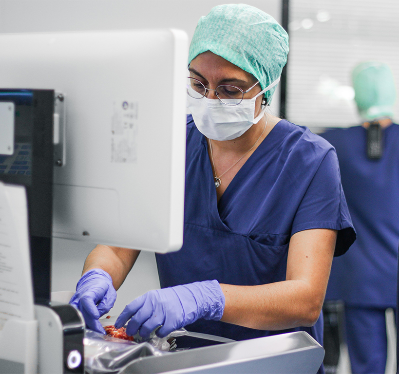
Dr Guani preparing to undertake a procedure
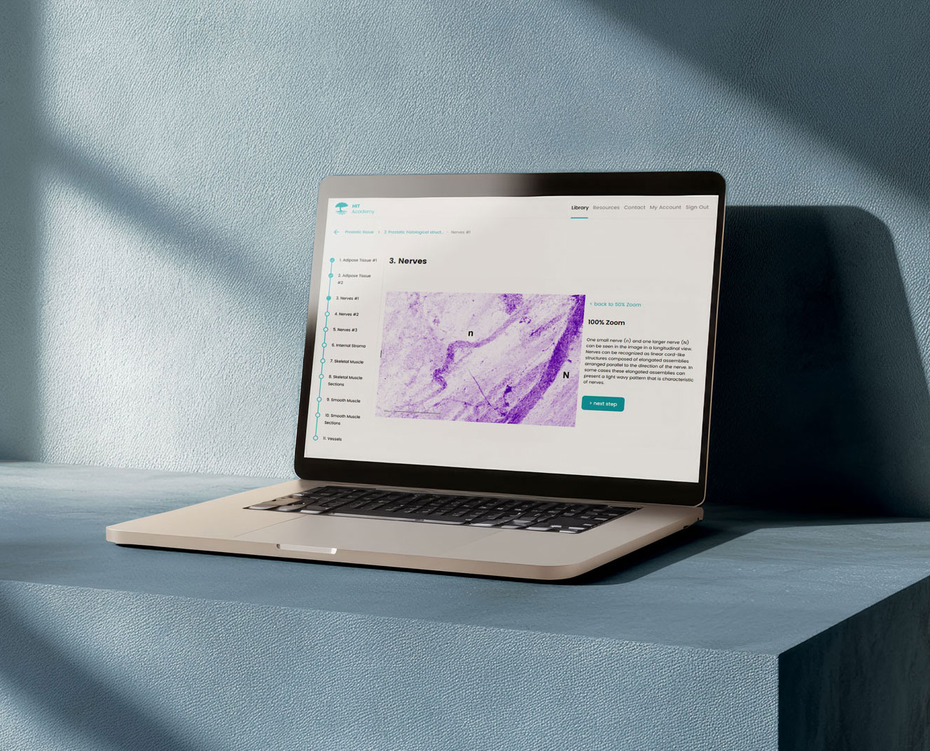
Histolog Image Training
The Histolog Image Training (HIT) was developed with our community of pathologists and experts to provide a simple and efficient way of getting familiar with Histolog images. Designed for both beginners and experienced morphology content readers, the HIT allows for flexible learning.
Curious about the Histolog Scanner ? Contact us.
A&P-00952 V1-International