The Histolog Scanner
Rapid imaging of tissue microstructures in excised specimens including cells, vessels and their organization or architecture.

Loading experience...
Rapid imaging of tissue microstructures in excised specimens including cells, vessels and their organization or architecture.
The Histolog Scanner integrates seamlessly into your workflow, with no calibration or setup required. It’s ready to use in minutes, delivering consistent, high-quality images without interruption.
The Histolog Scanner is the first and only massively parallel confocal microscopy device which allows the largest imaging field for issue imaging of 17cm² (4.8x3.6cm) with almost 30'000 micro-objectives. This enables physicians to visualize large fresh tissue samples in real time with outstanding resolution.
Once the tissue is available, simply dip the fresh tissue in Histolog Dip for 10 seconds, rinse it in saline and dry. Scanning takes about a minute per specimen surface, after which a physician can review and annotate images. The process is non-destructive, and does not affect the final pathology assessment.
Images are high-resolution and physicians can see cells, vessels and other tissue microstructures.
Easy navigation within the image, annotation of areas of interest, and more features.
Allowing users to easily access functions and operate the system.
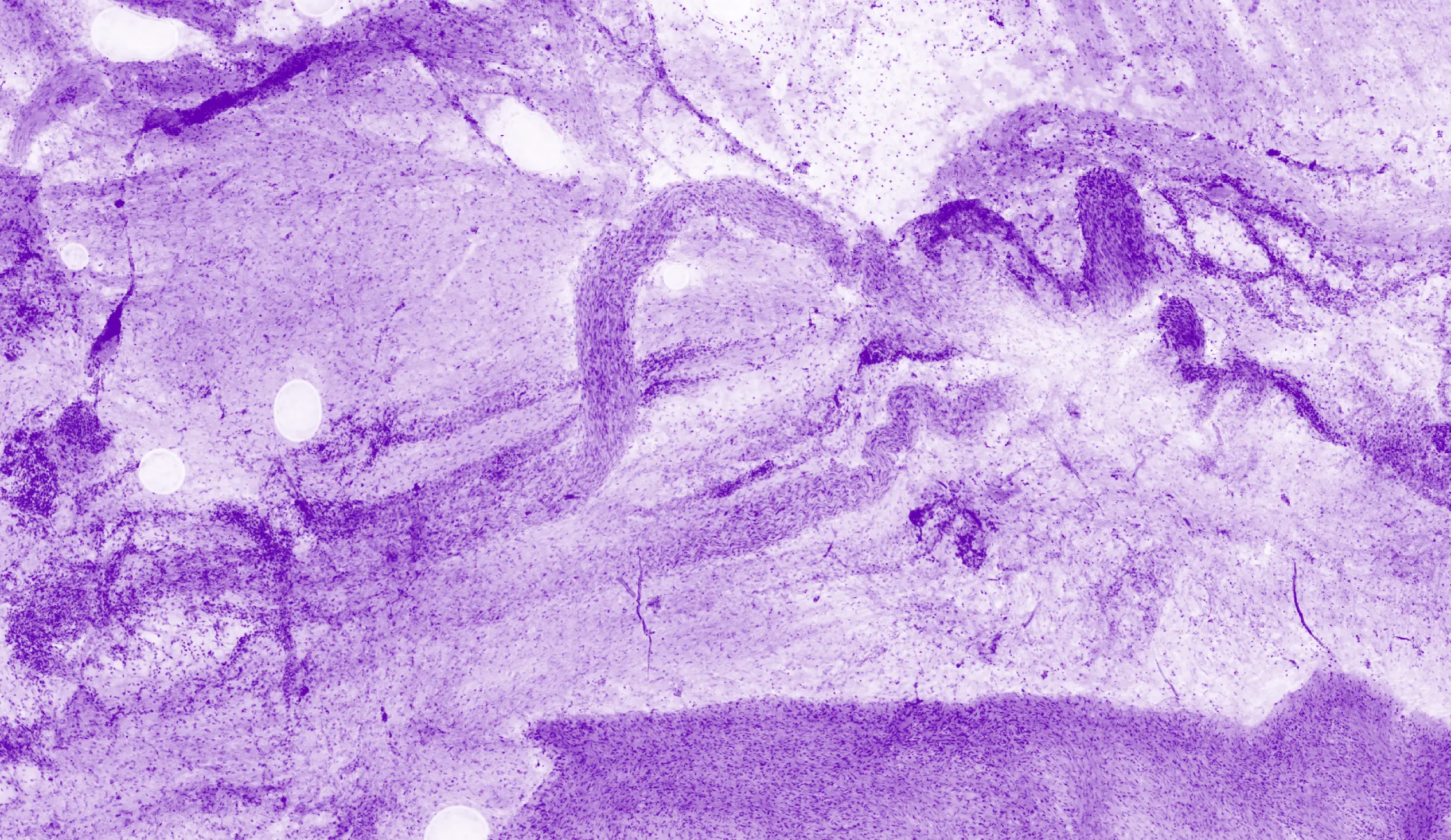
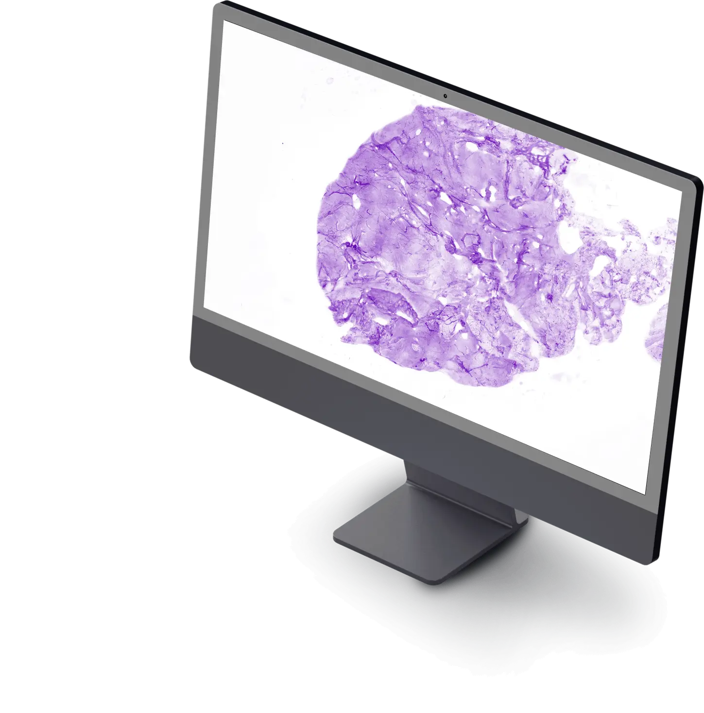
Histolog Remote enables physicians to access and review images from anywhere, supporting seamless collaboration and decision-making. It integrates into existing digital pathology workflows.
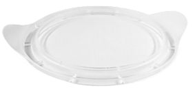
Designed with optical-grade imaging film to ensure high-resolution imaging, the Histolog Dish is a single-use, patented protection preventing direct contact of the tissue specimen with the Histolog Scanner during imaging.

The Histolog Dip is a single-use, proprietary fluorescent histological stain used when imaging fresh excised human tissue specimens with the Histolog Scanner. It is designed to preserve specimen integrity for future pathological analysis.
A&P-00951 V2-International
All data and images on file at SamanTree Medical.