

5-20 minutes
vs. 45-60 min with NeuroSAFE 1

79-91% sensitivity
Demonstrated across clinical studies 1,2,3

94-100% specificity
Demonstrated across clinical studies 1,2,3

Cost effective
Intraoperative assessment during RARP 4
References
1 Baas, D.J.H., Vreuls, W., Sedelaar, J.P.M. Vrijhof, H.J.E.J. Hoekstra, R.J., Zomer, S.F, van Leenders, G.J.L.H., Jvan Basten, J.A., Somford., D.M. Confocal laser microscopy for assessment of surgical margins
during radical prostatectomy. BJU Int. Published online 2022.
https://doi:10.1111/BJU.15938
2 Almeida-Magana R., et. al. Accuracy of the LaserSAFE technique for detecting positive surgical margins during robot-assisted radical prostatectomy: blind assessment and inter-rater agreement analysis. Histopathology. 2025 Feb;86(3):433-440.
https://doi: 10.1111/his.15336
3 Mayor, N., et al., IP8-FLUORESCE: A Prospective Paired Cohort Study Evaluating the Diagnostic Accuracy of Fluorescence Confocal Microscopy for Real-time Assessment of Surgical Margins in Radical Prostatectomy. Eur Urol, 2025.
https://doi.org/10.1111/bju.16588
4van Drumpt J.N.H.1, Govers T.M., Almeida-Magana R., Baas D.J.H.1, Haider A., Sedelaar J.P.M., Shaw G., Somford D.M. (2026). Cost-effectiveness of intraoperative surgical margin assessment during radical prostatectomy. BJU International 2026 https://doi:10.1111/bju.70121
Clinical workflow
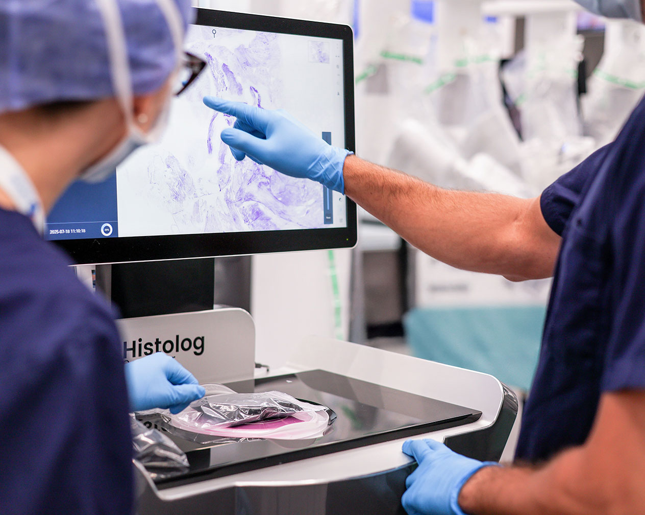
a 4-step procedure within 5 minutes
Excision
Collection of fresh tissue.
Preparation
Immerse the excision in Histolog Dip for 10 sec, and rinse it for 2 - 5 sec in saline. This process is non-destructive, requires no slicing and no slides.
Imaging
Scan the specimen surface in ~1 minute. The specimen remains visible and accessible during the entire procedure.
Evaluation
Review Histolog images immediately. This can be done on-site on the Histolog touchscreen, or remotely via Histolog Remote.
Focus on the results
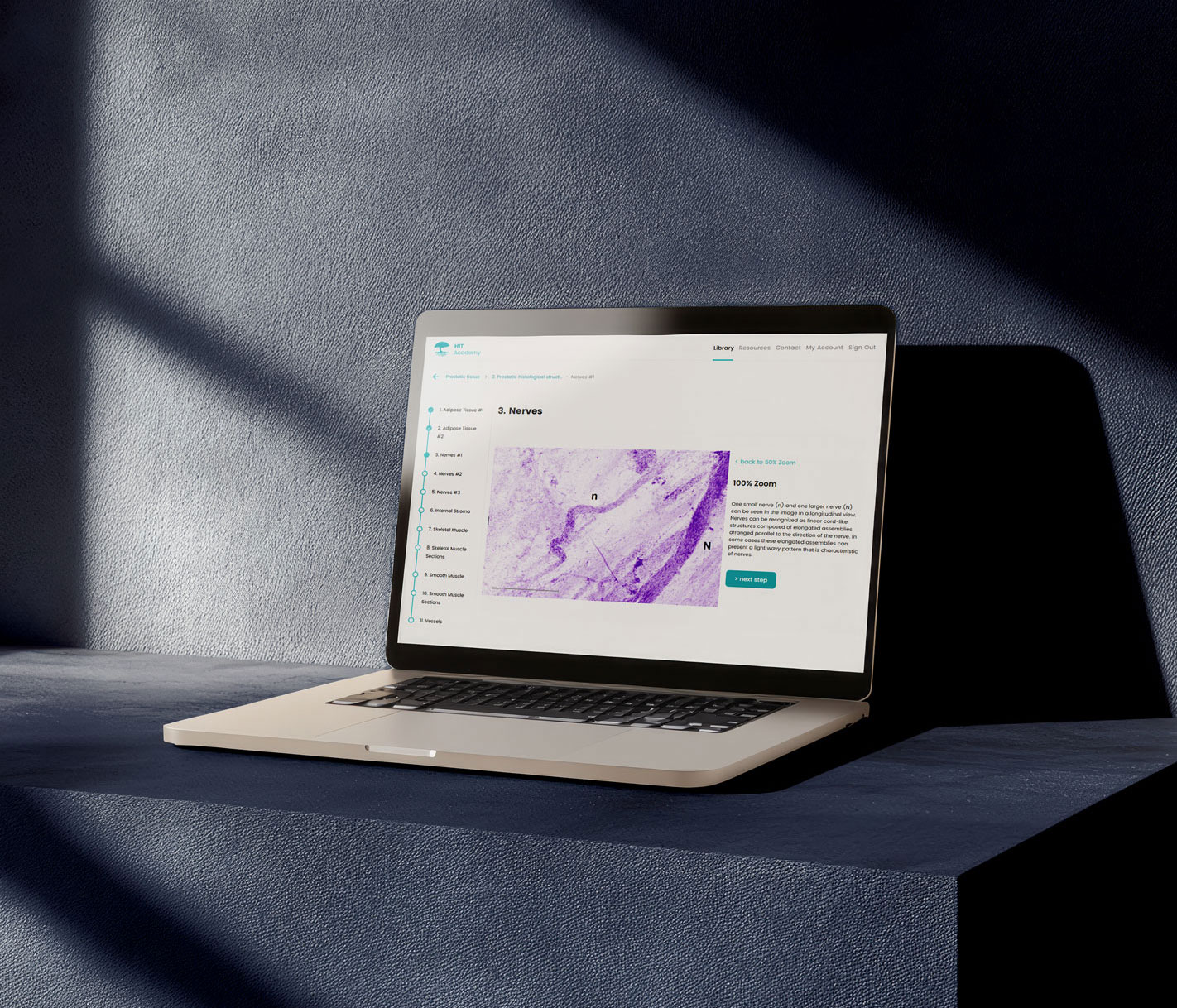
Histolog Image Training
The Histolog Image Training (HIT) was developed with our community of pathologists and experts to provide a simple and efficient way of getting familiar with Histolog images. Designed for both beginners and experienced morphology content readers, the HIT allows for flexible learning.
Curious about the Histolog Scanner ? Contact us.
A&P-00953 V2-International