Immediate margin assessment
Bringing the unseen into focus for a cancer-free tomorrow
From biopsy taking to surgical diagnosis, tissue assessment is a key component to efficient and straightforward cancer treatment. The challenge is to obtain high-resolution results in a timely manner.
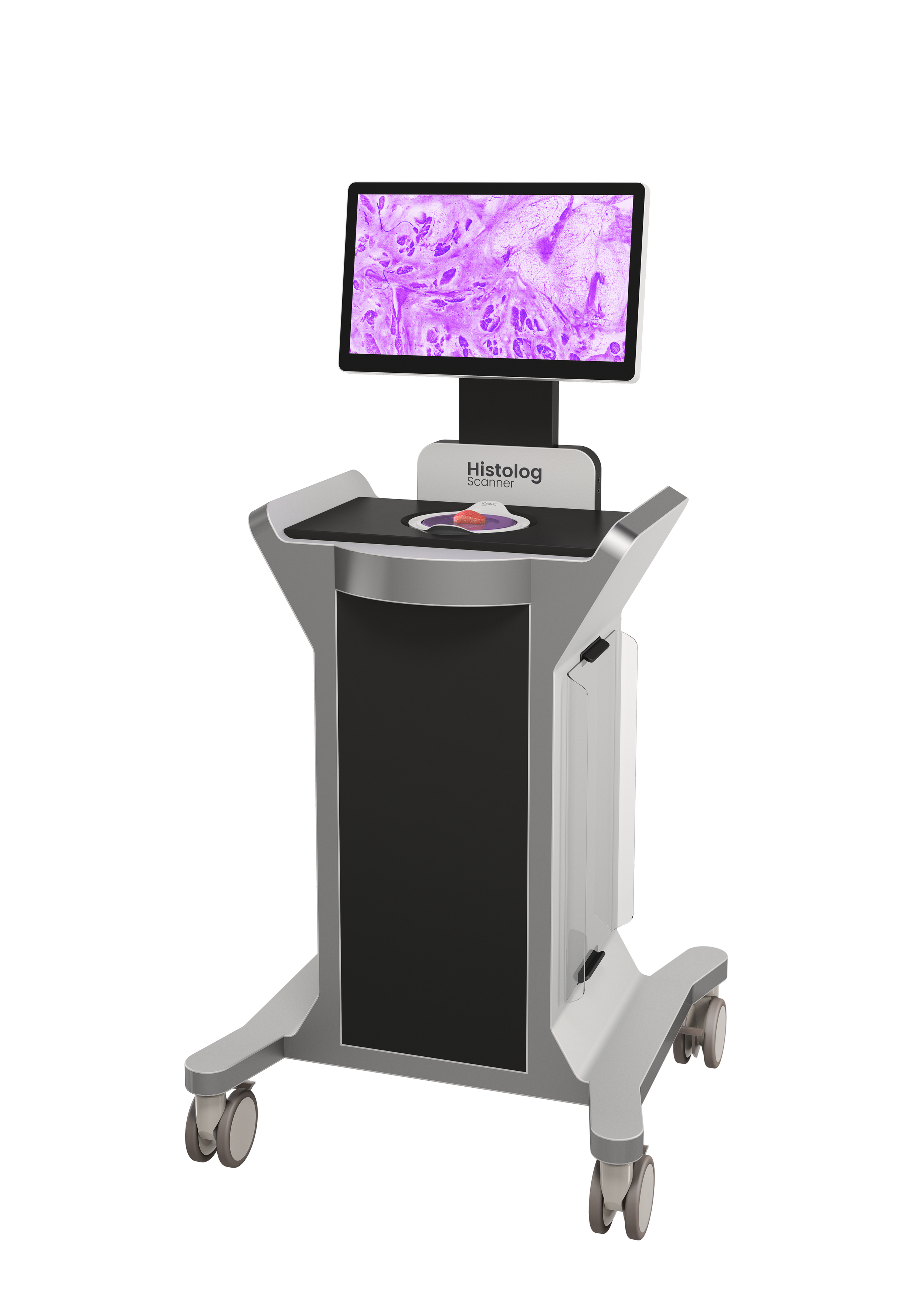
The Histolog® Scanner changes the game by bringing cancer cells to the fingertips of clinicians. It is a confocal microscopy device intended for imaging the surface of excised human tissue specimens to visualize morphological microstructures. Easy-to-read images are available immediately, providing decision-making support at the point of care. Start the video to see it in action.
Spot cancer cells when every minute counts
Device intended for qualified healthcare professionals only.
1.
Seamless clinical integration
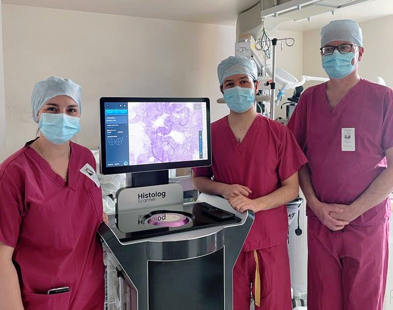
The Histolog Scanner is a plug-and-play device that sits perfectly in the operating theater or pathology laboratory.
2.
10-sec specimen preparation
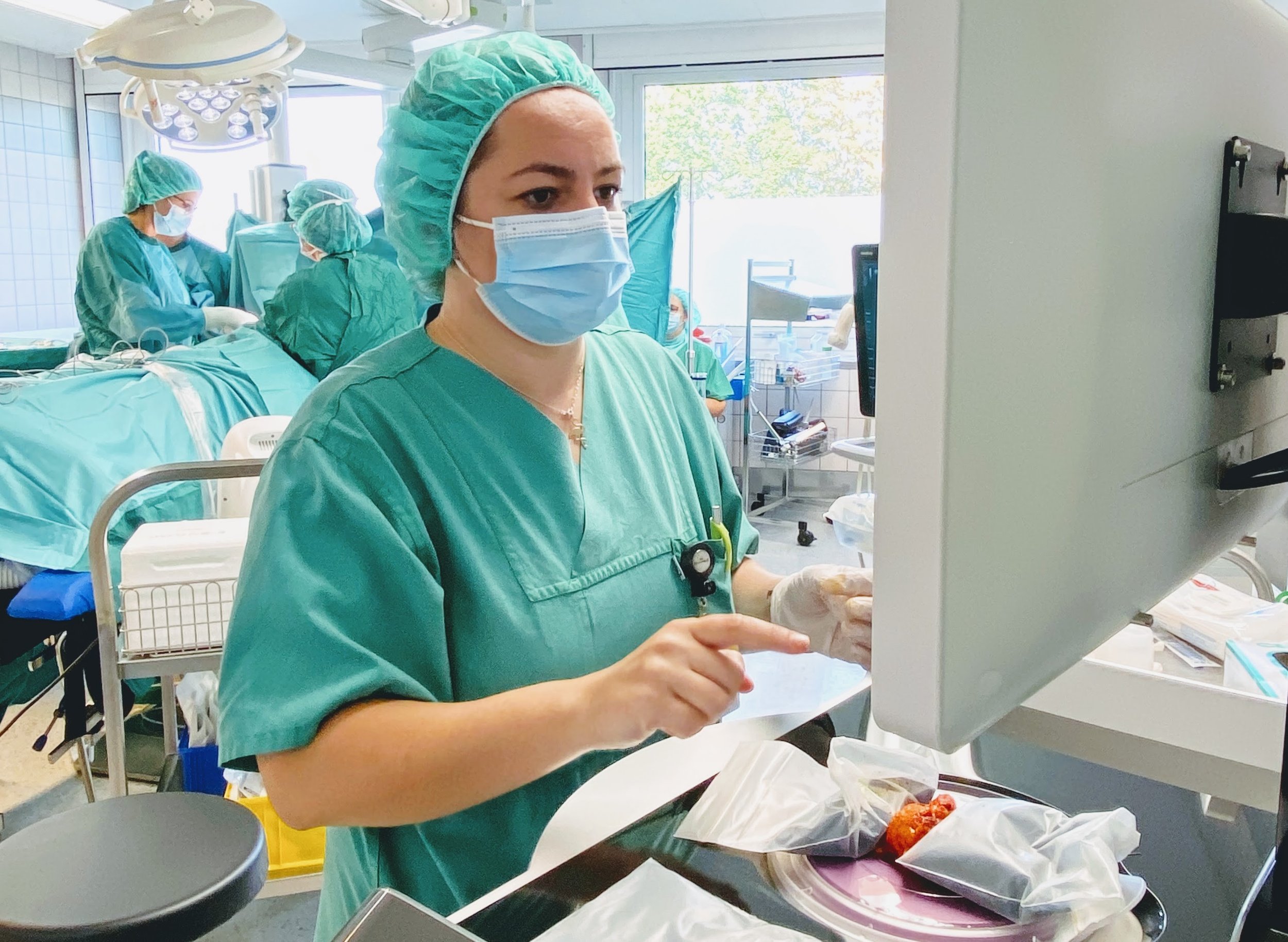
Once the specimen is resected, you only need to dip the fresh tissue for 10 sec in a fluorescent dye and rinse it before scanning.
3.
< 1 min to acquire an image
Scanning specimens up to 〜17cm² takes less than a minute. Easy-to-read images of the whole surface area are available rapidly to make sure that no cancer cell remains.
4.
Remote workflow in a single tap
You can work remotely with fellow doctors or pathologists thanks to the Histolog Digital Solution, which works towards easy storing and sharing of images.
The Histolog® Scanner
The Histolog Scanner (CE mark since 2018) is a digital microscopy scanner for high-resolution imaging of the surface of fresh tissue. It is based on a novel ultra-fast confocal microscopy technology invented in EPFL in 2010. Its innovative design makes it highly practical for quick assessment on the point-of-care and the clinician is one touch on the screen away from visualizing cancerous cells immediately on a surgical specimen ¹ ⁻ ⁸.
The Histolog Dip and Dish
The Histolog Dip (CE-IVD) is a fluorescent histological stain intended to be used as an accessory device in combination with the Histolog Scanner for imaging the surface of excised human tissue specimens to visualize morphological microstructures. It is designed for rapid imaging of fresh tissue, while preserving specimen integrity for future pathological analysis ² ⁻ ⁵.
The Histolog Dish (CE-IVD) is a single-use protection preventing direct contact of the tissue specimen with the Histolog Scanner during imaging.
The Histolog Viewer
The Histolog Viewer is the image management system software intended to be used with the Histolog Scanner. Designed to help you quickly and easily examine specimens morphological structures, this powerful software allows you to access, display, annotate, manage, store, and share digital Histolog Scanner images and metadata. Plus, with advanced capabilities for further analyzing images and marking regions of interest (ROI), the Histolog Viewer takes your capabilities to the next level (ROI management feature for breast).
Request an e-demo
Experience the Histolog Scanner firsthand and discover how it can transform your workflow.
Proven in prostate, breast and beyond
CLINICAL STUDIES
-
Comparison of the Histolog Scanner approach to NeuroSAFE procedure, Prof. D. Somford, Canisius Wilhelmina Hospital, Nijmegen – Netherlands BJUI 2022
- Similar performance as NeuroSAFE
- Drastic time saving : 80% time-reduction (average 8 min vs. 50 min)
- Low resources required : used in autonomy by pathologistResearch letter - Improving fluorescence confocal microscopy for margin assessment during robot-assisted radical prostatectomy: The LaserSAFE technique, Prof G. Shaw, UCLH, London – UK BJUI 2023
-
ATLAS STUDIES
BCS Atlas: Breast Tissue imaging Atlas using Ultra-fast Confocal Microscopy to identify cancer lesions, Dr. Mathieu, Institut Gustave Roussy – France, Virchows Archiv 2024
Core Needle Biopsies: Comparative analysis of confocal microscopy on fresh breast core needle biopsies and conventional histology, Prof. Tausch - Switzerland, Diagnostic Pathology 2019
INITIAL STUDIES
Breast carcinoma detection in ex vivo fresh human breast surgical specimens using a fast slide-free confocal microscopy scanner: HIBISCUSS project, Angelica Conversano, Gustave-Roussy Institute, France, BJS Open 2023
High performances for both pathologists (Sensitivity >95%, Specificity >95%) and surgeons (Sensitivity >90%, Specificity >90%)
Short learning curve to understand Histolog image content
Superiority to standard-of-care, potential reduction of re-operation up to 75%- Detection of DCIS on the surface AND in-depth
Compatibility with clinical routine (2 min average usage per margin)
Development of Histolog Image Training program
Intraoperative insertion of Histolog Scanner and initial performance of clinicians, Prof. M. Golatta, Heidelberg University Hospital – Germany, The Breast 2023
NEXT GEN. STUDIES SUPPORTED BY THE HIT ACADEMY
Real-Time Scanning of Fresh Lumpectomy with ‘Histolog Digital Solution’ for Breast Conserving Surgery: Routine Utilization Study, Prof. M. P. Lux, St. Vincenz Hospital – Germany, ongoing
Insertion into clinical routine, remote assessment by pathologist, Dr. M. Kholik, Genolier Clinic – Switzerland, ongoing
-
First insertion into Mohs surgery setting, Prof. M. Möhrle, University of Tübingen – Germany JEADV 2018
- Time-saving and very effective alternative to classical paraffin-embedded or frozen section
- BCC detection performance: sensitivity >70% and specificity 96%Performances of the Histolog Scanner in Mohs surgery, Dr. F. Kuonen, CHUV – Switzerland Skin and Health Disease 2022
- Drastic time saving
- Basal Cell Carcinoma: Sensitivity >80% and specificity 100%
- Realistic approach for Mohs surgery
- Development of dermatology AtlasEx vivo confocal microscopy for the intraoperative assessment of deep margins in giant basal cell carcinoma, Dr. F. Kuonen, CHUV – Switzerland JAAD case report 2022
- Successful scanning and identification of cancer lesions with the Histolog Scanner
- Make IOMA for giant specimens possible
- Allow direct reconstruction
-
2021, Imaging evaluation of multiple tissues and assessment of benefits for pathology lab, Prof. F. v. Kemenade, Erasmus Hospital Pathology Department, Erasmus – Netherlands
2022, Evaluation of the Histolog Scanner in neurosurgery, Mr. R. Mathew, Leeds Teaching Hospitals NHS Trust, Leeds – United Kingdom
2022, Evaluation of the Histolog Scanner in ENT cancer, Dr. JP. Jeanon, Guy’s hospital King’s University, London – United Kingdom

Histolog Image Training
Only a few hours to learn how to read Histolog images
The Histolog Image Training (HIT) was developed with our community of pathologists and experts to provide a simple and efficient way of getting familiar with Histolog images. Designed for both beginners and experienced morphology content readers, the HIT is accessible to all and allows for flexible learning. You will be ready to interpret morphology images in just a few hours of training.


